Scanner may be extraordinary in breast cancer fight
BBC Scotland, Aberdeen

 University of Aberdeen
A scanner developed in Aberdeen could be potentially “extraordinary” for diagnosing and treating breast cancer, it has been claimed.
Scientists from the University of Aberdeen used a prototype version of the new Field Cycling Imager (FCI) scanner to examine the breast tissue of patients newly-diagnosed with cancer.
They said they found that the FCI scanner could distinguish tumour material from healthy tissue with more accuracy than current Magnetic Resonance Imaging (MRI) methods.
While similar to MRI – which was also developed in Aberdeen – the FCI scanner is described as being able to vary the strength of the magnetic field during the patient’s scan.
The university described the scanner as a “world first”, which could identify “previously undetectable cancer tumour invasion”.
The scientists worked in collaboration with NHS Grampian on the study.
The technology being able to detect tumours without having to inject dye into the body was described as a further benefit.
The team said the success with breast tissue followed earlier positive outcomes when the prototype was used to identify brain damage due to a stroke.
‘Potential is limitless’
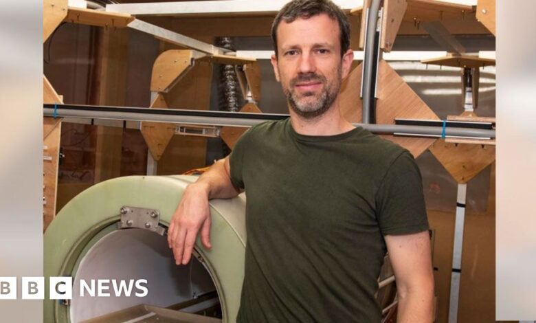
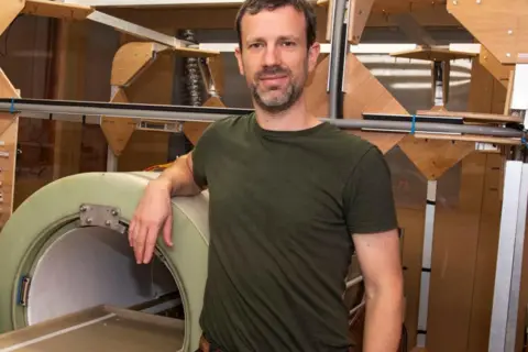
Dr Lionel Broche is a senior research fellow in biomedical physics and the lead researcher in the study.
“We found that images generated from FCI can characterise breast tumours more accurately,” he said.
“This means it could improve the treatment plan for the patients by improving the accuracy of biopsy procedures by better detecting the type and location of tumours, and by reducing repeated surgery so really, the potential impact of this on patients is extraordinary.”
He added: “My colleagues in the University of Aberdeen built the world’s first clinical MRI in the 1970s so it is both fitting and exciting that we are making waves again with an entirely new type of MRI called FCI.
“This is a truly exciting innovation and as we keep improving the technology for FCI, the potential for clinical applications is limitless.”
Dr Gerald Lip is a consultant radiologist in NHS Grampian and co-investigator in the study, who has recently been appointed president of the British Society of Breast Radiology.
“This data is very promising, and we still need more prospective work, but these results will really support future clinical applications,” he said.
“We treat between 400 and 500 women with breast cancer in NHS Grampian every year and the potential this technology has to reduce the need for women to return for extra surgery is huge, benefiting them and reducing wait times and operating theatre resource.
“We hope it will have a future role in supporting cancer diagnosis and management.”